
Intestinal Ultrasonography
What is Intestinal Ultrasonography?
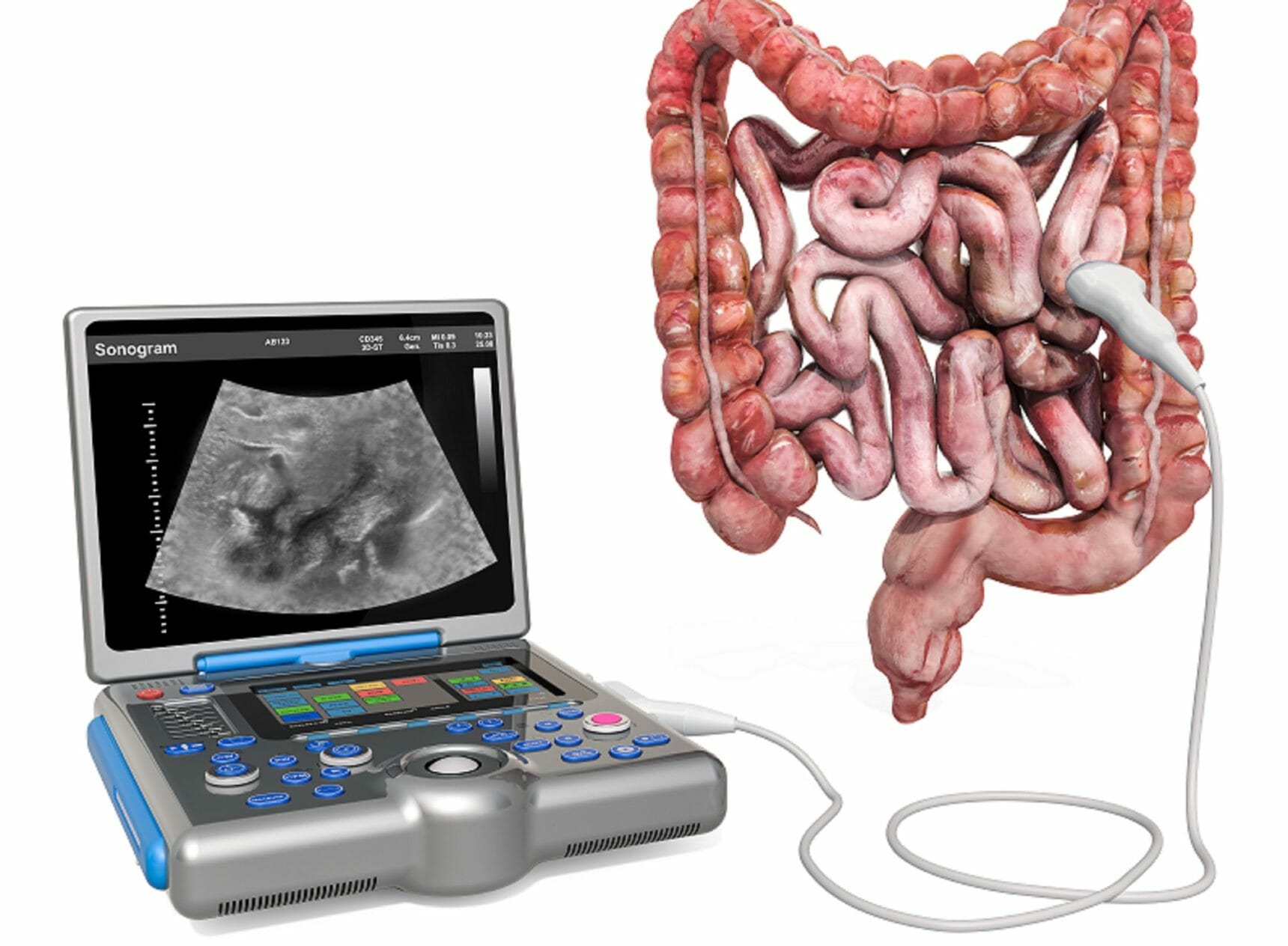
Intestinal Ultrasonography is a non-invasive imaging technique used to examine the intestines and surrounding abdominal structures. It uses high-frequency sound waves to create detailed images of the small and large intestines, helping doctors detect abnormalities such as inflammation, thickening, obstructions, or masses.
Unlike other imaging methods that require radiation, intestinal ultrasonography is safe, painless, and radiation-free, making it particularly suitable for children, adolescents, and adults with chronic intestinal conditions.
Dr. Harshad V. Joshi, a leading Gastroenterologist in Mumbai, specializes in performing intestinal ultrasonography with precision, providing accurate diagnoses and tailored treatment plans.
Why is Intestinal Ultrasonography Important?
Intestinal ultrasonography is a critical diagnostic tool in gastroenterology for several reasons:
- Early Detection: Identifies issues like Crohn’s disease, ulcerative colitis, tumors, and intestinal obstructions at an early stage.
- Monitoring Chronic Conditions: Helps track disease progression in patients with Inflammatory Bowel Disease (IBD) or other chronic intestinal disorders.
- Guiding Treatment: Provides detailed images that allow doctors to plan appropriate medical or surgical interventions.
- Non-Invasive and Safe: No radiation exposure, making it suitable for frequent monitoring.
Conditions Diagnosed with Intestinal Ultrasonography
Intestinal ultrasonography can help detect and monitor a wide range of conditions, including:
- Inflammatory Bowel Disease (IBD): Crohn’s disease and ulcerative colitis can be monitored effectively, assessing inflammation and bowel wall thickening.
- Intestinal Obstructions: Detects blockages caused by adhesions, tumors, or strictures.
- Tumors and Polyps: Early identification of abnormal growths in the small and large intestines.
- Diverticulitis: Detects inflamed or infected pouches in the colon wall.
- Appendicitis: Helps in confirming suspected cases of appendiceal inflammation.
- Vascular Abnormalities: Detects issues related to blood flow in the intestines.
Advantages of Intestinal Ultrasonography
- Painless and Comfortable: No needles or radiation; the procedure is quick and minimally invasive.
- Real-Time Imaging: Provides live images of intestinal movement and blood flow.
- Cost-Effective: Less expensive than CT scans or MRI, yet highly effective for initial evaluation.
- Frequent Monitoring: Can be repeated multiple times for ongoing assessment without risk.
What to Expect During the Procedure
- Preparation – Patients may be asked to fast for a few hours prior to the scan.
- Positioning – The patient lies on an examination table.
- Ultrasound Application – A gel is applied to the abdomen to improve image quality.
- Scanning – The gastroenterologist moves the ultrasound probe over the abdomen to capture images of the intestines.
- Result Analysis – Images are analyzed in real-time, and findings are discussed with the patient.
The entire process is usually completed within 30–45 minutes, and patients can resume normal activities immediately after.
Book Your Session Today
If you are experiencing persistent abdominal pain, unexplained digestive symptoms, or need regular monitoring for a gastrointestinal condition, Intestinal Ultrasonography could be the right diagnostic tool for you.
Schedule your appointment today with Dr. Harshad V. Joshi – Gastroenterologist in Mumbai, and get accurate, safe, and reliable intestinal health evaluation.
Book an AppointmentFrequently Asked Questions (FAQs)
Yes, it is completely safe, non-invasive, and free from radiation, making it ideal for young patients.
You may be asked to fast for 4–6 hours before the procedure and avoid carbonated drinks for better imaging.
Yes, it can identify suspicious masses and abnormalities that may indicate early stages of cancer, but additional tests may be required for confirmation.
No, intestinal ultrasonography is painless. You may feel slight pressure as the probe moves over the abdomen.
Frequency depends on the condition being monitored. Chronic conditions like Crohn’s disease may require periodic scans for assessment and management