
Enteroscopy in Mumbai
Enteroscopy
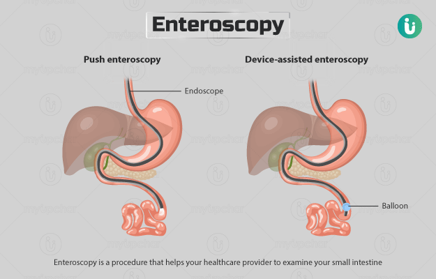
Enteroscopy is a medical procedure used to visualize and examine the small intestine, which is located between the stomach and the large intestine (colon). The small intestine is relatively difficult to access using traditional imaging methods like endoscopy or colonoscopy due to its length and location. Enteroscopy allows healthcare providers to directly examine the mucosal lining of the small intestine and diagnose or treat various gastrointestinal conditions.
There are two main types of enteroscopy:
- 1. Single-Balloon Enteroscopy: In this technique, a specialized endoscope with a balloon at its tip is inserted through the mouth or anus. The balloon can be inflated to anchor the scope, allowing it to be advanced deep into the small intestine. This type of enteroscopy is useful for both diagnostic purposes and therapeutic interventions. If you are looking for advanced Balloon Enteroscopy in Mumbai, this procedure is one of the commonly preferred methods.
2. Double-Balloon Enteroscopy: This method employs an endoscope with two balloons, one at the tip and another on the side. The balloons are alternately inflated and deflated to advance the scope through the small intestine in a “step-by-step” manner. This approach provides even greater reach into the small intestine and allows for more comprehensive examination.
3. Spiral motorized enteroscopy: Spiral motorized enteroscopy, also known as spiral enteroscopy or spiral overtube-assisted enteroscopy, is a specialized technique used to examine the small intestine’s inner lining in a comprehensive and controlled manner. This procedure is designed to overcome some of the challenges associated with traditional endoscopic methods, which can be limited by the small intestine’s length and complex structure.
- 1. Single-Balloon Enteroscopy: In this technique, a specialized endoscope with a balloon at its tip is inserted through the mouth or anus. The balloon can be inflated to anchor the scope, allowing it to be advanced deep into the small intestine. This type of enteroscopy is useful for both diagnostic purposes and therapeutic interventions. If you are looking for advanced Balloon Enteroscopy in Mumbai, this procedure is one of the commonly preferred methods.
Enteroscopy is used for several purposes:
- Diagnosis: It can help diagnose various conditions affecting the small intestine, including Crohn’s disease, tumors, ulcers, bleeding, celiac disease, and other inflammatory or structural disorders.
- Biopsies: During enteroscopy, tissue samples (biopsies) can be taken from the small intestine’s lining to aid in diagnosing certain diseases.
- Polyp and Lesion Removal: If polyps, lesions, or abnormal growths are identified, enteroscopy can be used to remove or treat them.
- Bleeding Management: Enteroscopy can help identify the source of gastrointestinal bleeding in the small intestine and potentially allow for interventions to stop the bleeding.
Enteroscopy is typically performed with the patient under sedation or anesthesia to ensure comfort during the procedure. Like other endoscopic procedures, it carries some risks, such as bleeding, infection, and perforation, although these risks are generally low. The preparation for enteroscopy can involve dietary restrictions and bowel cleansing to ensure clear visibility of the small intestine’s lining.
The specific type of enteroscopy and the reason for the procedure will determine whether it is performed through the mouth (oral) or through the anus (anal). Your healthcare provider will guide you on the appropriate type of enteroscopy and the necessary preparations based on your medical history and symptoms.
Double-balloon enteroscopy (DBE) is a specialized endoscopic procedure used to visualize and examine the small intestine, which is otherwise challenging to access with traditional endoscopy or colonoscopy due to its length and convoluted structure. DBE employs a unique technique involving two balloons—one at the tip of the endoscope and another on the side—to help advance the scope through the small intestine in a step-by-step manner.
Here's how double-balloon enteroscopy works:
- Insertion: The endoscope, which has two balloons attached, is inserted through the mouth or anus, similar to other endoscopic procedures. The first balloon is inflated at the tip of the scope.
- Balloon Inflation: The inflated tip balloon helps anchor the scope in place, allowing the second balloon (side balloon) to be inflated further along the scope.
- Advancement: The side balloon is inflated and deflated sequentially, creating a “push-and-pull” effect that propels the endoscope forward or backward in the small intestine. This controlled inflation and deflation allow the scope to be moved through the intestine in a controlled manner.
- Visualization and Examination: As the endoscope is advanced, the camera at its tip transmits images to a monitor, providing real-time visualization of the small intestine’s lining. The healthcare provider can examine the mucosa, identify abnormalities, and perform procedures if needed.
- Step-by-Step Approach: The scope is maneuvered through the small intestine incrementally, making it possible to explore the entire length of the small intestine in a more thorough manner compared to other methods.
Double-balloon enteroscopy is used for various purposes, including:
- Diagnosis: It can aid in diagnosing conditions affecting the small intestine, such as Crohn’s disease, small intestinal tumors, ulcers, and other inflammatory or structural disorders.
- Biopsies: Tissue samples (biopsies) can be taken during the procedure to help diagnose specific diseases or conditions.
- Treatment: If polyps, growths, or other abnormalities are found, they can be removed or treated using specialized tools attached to the endoscope.
Double-balloon enteroscopy is typically performed with the patient under sedation or anesthesia to ensure comfort and minimize discomfort during the procedure. As with any endoscopic procedure, there are potential risks, such as bleeding, infection, or perforation, although these risks are generally low. Your healthcare provider will provide you with specific instructions for preparation and recovery based on your individual situation and medical history.
Spiral enteroscopy: In spiral motorized enteroscopy, a specialized overtube is used in conjunction with the endoscope to achieve better access and visualization of the small intestine. The overtube is a flexible tube that surrounds the endoscope and can be advanced through the intestine, providing stability and facilitating advancement.
Here's how spiral motorized enteroscopy works:
- Insertion: The overtube and endoscope assembly are inserted either through the mouth (oral) or the anus (anal), depending on the approach chosen by the healthcare provider. The overtube’s design allows it to be advanced deeper into the small intestine.
- Spiral Movement: Unlike traditional endoscopy, where the scope is pushed and pulled in a linear manner, spiral motorized enteroscopy involves a rotating movement. The overtube and endoscope are rotated in a spiral manner, allowing for more effective advancement through the twists and turns of the small intestine.
- Visualization and Examination: As the overtube and endoscope are rotated and advanced, the camera at the endoscope’s tip transmits images to a monitor, providing a real-time view of the small intestine’s mucosal lining. The healthcare provider can examine the intestinal walls, identify abnormalities, and perform interventions if necessary.
- Comprehensive Coverage: The spiral movement of the overtube and endoscope enables more thorough exploration of the small intestine’s length, potentially reaching areas that might be difficult to access using other methods.
Spiral motorized enteroscopy serves similar purposes to other enteroscopy techniques, including diagnosing conditions, taking biopsies, removing polyps or growths, and treating bleeding or other abnormalities within the small intestine.
This technique aims to provide a more efficient and thorough examination of the small intestine while minimizing patient discomfort. As with any endoscopic procedure, there are potential risks and complications, such as bleeding, infection, or perforation, although these risks are generally low. Patients will receive instructions from their healthcare provider regarding preparation and recovery specific to the spiral motorized enteroscopy procedure.